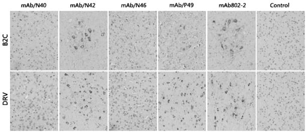
Fig. 3.
Characterization of anti-N Mabs in immonuhistochemical analysis. RABV-infected brain tissues were fixed in formalin and embedded in paraffin. Coronal sections were prepared and reacted with each of the Mabs, followed by biotinylated secondary antibody (goat anti-mouse) and avidin-peroxidase. Finally, diaminobenzidine (DAB) was used as a substrate for color development. B2C brain tissues from mice infected with RABV B2C. DRV brain tissues from mice infected with RABV B2C

