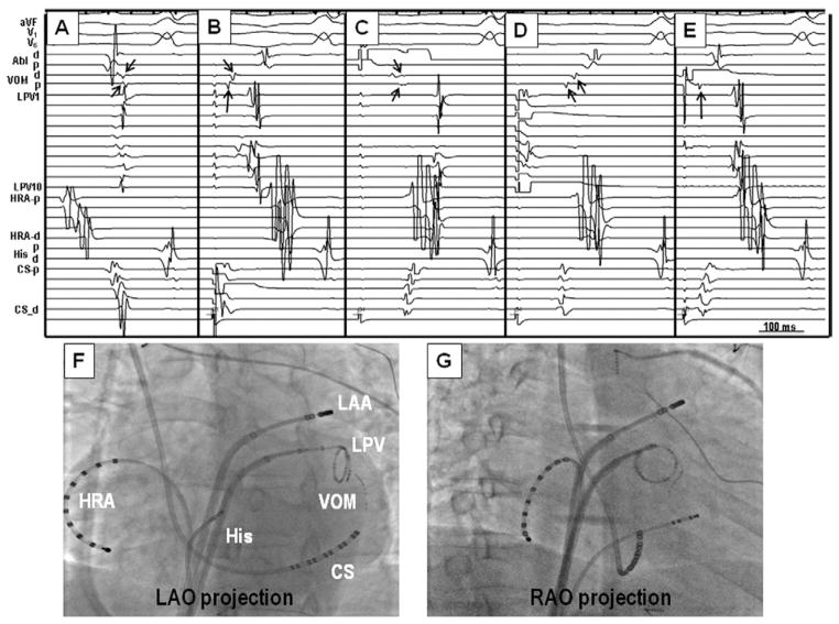
Figure 4.
Example of differential pacing to map the MP potentials. A: Intracardiac recordings during sinus rhythm. B: Activation during CS pacing. The earliest activation was in CS, followed by proximal-to-distal propagation within the VOM. These VOM potentials (arrows) clearly preceded the onset of PV and HRA potentials. C: Pacing from distal pole of the ablation catheter located within the LAA. The potentials recorded by the VOM catheter (arrows) are completely separated from atrial and PV potentials. D: Pacing from left PV separated the VOM potentials (arrows) from the PV potentials. E: Pacing from the distal VOM showed that the proximal VOM potential (arrow) preceded the PV potential, indicating that these potentials are not from the PVs. F and G: Fluoroscopic images during differential pacing. Abl = ablation; HRA = high right atrium; LAA = left atrial appendage; LAO = left anterior oblique; LPV = left pulmonary vein; RAO = right anterior oblique; other abbreviations as in Figure 3.

