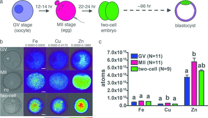
Figure 1. Synchrotron-based x-ray fluorescence microscopy reveals intracellular distribution of the transition elements in oocytes and early embryo.
Fully-grown oocytes, eggs, and two-cell embryos (a) were prepared as whole mount samples for synchrotron-based xray fluorescence microscopy. Oocytes (N = 11) display an intact germinal vesicle (GV, asterisk) while mature (MII) eggs (N = 11) have a visible first polar body (PB). Two-cell embryos (N = 9) were obtained by in vitro fertilization. Representative brightfield images for each stage are shown, in addition to the elemental maps for iron, copper and zinc (b). The minimum and maximum elemental content (μg/cm2) are shown above each set of elements. Among the biologically relevant transition elements, zinc is an order of magnitude more abundant than iron and copper at all developmental stages (c). Scale bar = 20 μm. Data represent mean values ± s.e. Letters denote statistically significant differences between developmental stages for individual elements (p<0.05).

