Fig. 1.
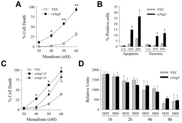
Inhibition of macroautophagy sensitizes to death from menadione. (A) VEC and siAtg5 cells were treated with the indicated menadione concentrations for 24 h and the percentage of cell death determined by MTT assay (*P<0.001 and **P<0.000001 as compared to VEC cells treated with the same menadione concentration; n=7). (B) VEC and siAtg5 cells were treated with 50 (M50) or 60 (M60) μM menadione for 12 h, costained with acridine orange and ethidium bromide, and the percentages of apoptotic and necrotic cells determined by fluorescence microscopy as described in Materials and Methods (*P<0.0001 as compared to the same treatment in VEC cells; n=6–7). (C) Percentage of cell death by MTT assay in VEC, siAtg5 #2 and siAtg5 #3 cells after 24 h of treatment with the indicated concentrations of menadione (*P<0.0001 as compared to the same treatment in VEC cells; n=6–8). (D) Relative levels of ROS in the two cell types at different times after treatment with 50 or 60 μM menadione (n=3–6).

