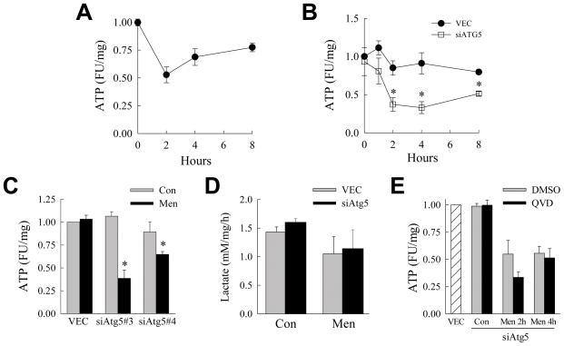
Fig. 5.
Inhibition of macroautophagy leads to ATP depletion in response to menadione. (A) Relative ATP levels in fluorescence units (FU) in VEC cells treated with 60 μM menadione for the indicated times (n=5–8). (B) ATP levels in VEC and siAtg5 cells treated with 50 μM menadione for the times shown (*P<0.01 as compared to VEC cells; n=3–4). (C) ATP levels in VEC, siAtg5 #2 and siAtg5 #3 cells untreated (Con) and after 2 h of 50 μM menadione (Men) (*P<0.03 as compared to untreated cells; n=4–6). (D) Rates of lactate production in VEC and siAtg5 cells untreated and treated with 50 μM menadione for 2 h (n=3). (E) Levels of ATP in untreated VEC cells and siAtg5 cells untreated (Con) or treated with 50 μM menadione for 2 or 4 h after pretreatment with dimethylsulfoxide (DMSO) or Q-VD-OPh (QVD) ( n=4).

