Fig. 7.
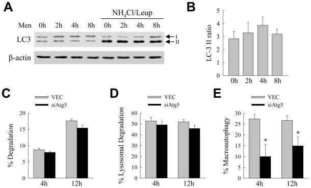
Other forms of autophagy are up regulated in compensation for the loss of macroautophagy. (A) Immunoblots of proteins isolated from VEC cells untreated and treated with menadione for the times shown in the absence or presence of ammonium chloride/leupeptin (NH4Cl/Leup) and probed for LC3 and β-actin. Ammonium chloride/leupeptin was added to the cells 2 h prior to isolation. The LC3-I and LC3-II bands are indicated by arrows. (B) Ratio of the signal intensity of the LC3-II bands in the presence of inhibitors to that in the absence of inhibitors as determined by densitometric scanning of 3 immunoblots. (C) Percentage of protein degradation in VEC and siAtg5 cells at the indicated times as measured by metabolic pulse-chase labeling (n=4). (D) Percentage of lysosomal degradation in the same cells as determined by the amount of protein degradation that was inhibited by ammonium chloride/leupeptin (n=4) (E) Percentage of lysosomal degradation that was mediated by macroautophagy which was the amount inhibited by 3-methyladenine (*P<0.05 when compared to VEC cells; n=4).

