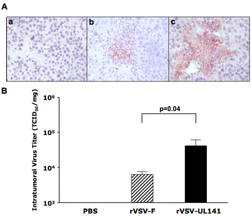
Figure 3. rVSV-UL141 versus rVSV-F replication in HCC tumors in the livers of immune-competent Buffalo rats.

Multi-focal HCC-bearing Buffalo rats were treated with PBS (n=3), rVSV-F (n=4) or rVSV-UL141 (n=4) at 1×10⁷ pfu/rat injected through the hepatic artery. Tumor samples were obtained from the treated rats at day 3 after virus infusion. 5 µm tumor sections were stained with a monoclonal anti-VSV-G antibody and counterstained with Hematoxylin (Panel A). Representative sections from rats treated with PBS, rVSV-F and rVSV-UL141 are shown in frames a, b and c, respectively (magnification=40×). In panel B, intratumoral virus titers were determined by TCID50 assays performed using tumor extracts on BHK-21 cells. Viral titers are expressed as TCID50/mg tissue (mean ± sstandard deviation), and the results were analyzed statistically by two-sided student t test.
