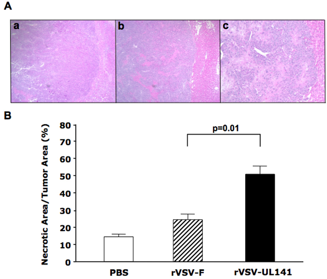
Figure 4. Enhanced tumor response in rats treated with rVSV-UL141 versus those treated with rVSV-F.

Multi-focal HCC-bearing Buffalo rats were injected via the hepatic artery with PBS (n=3), rVSV-F (n=4) or rVSV-UL141 (n=4) at 1×10⁷ pfu/rat and sacrificed 3 days post-treatment. Panel A, 5 µm tumor sections were stained with H&E. Representative sections from rats treated with PBS, rVSV-F and rVSV-UL141 are shown in frames a, b and c, respectively (magnification=40×). Panel B, Percentage of necrotic areas in the tumors were measured morphometrically using ImagePro software. Data are shown as mean ± standard deviation, and the results were analyzed statistically by two-sided student t test.
