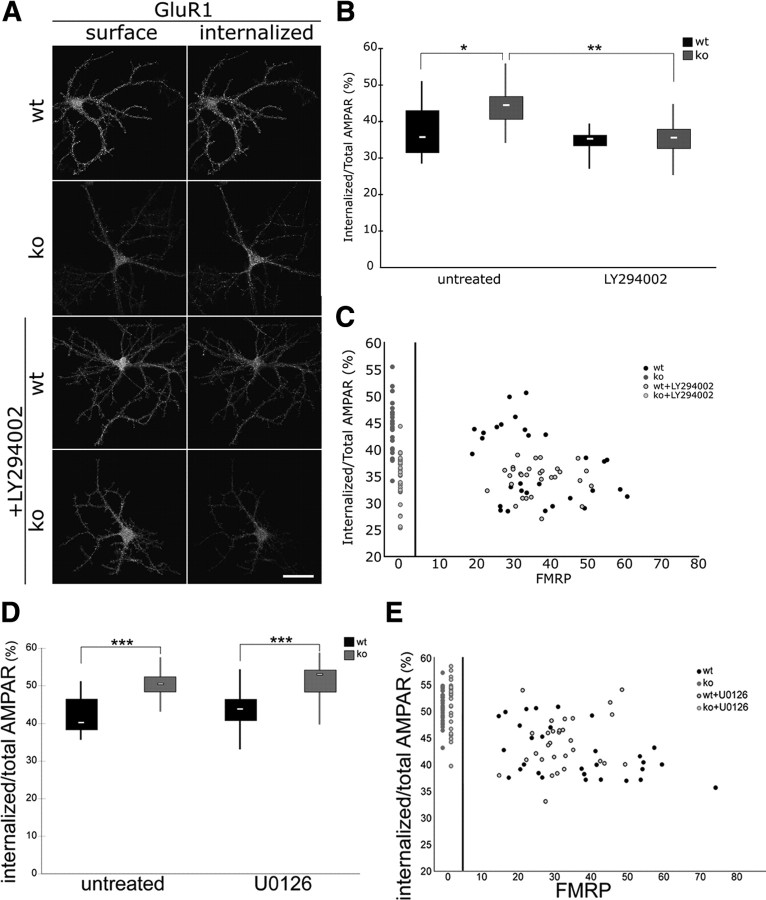
Figure 6.
A PI3K antagonist rescues increased GluR1 endocytosis in Fmr1 KO neurons. A, Representative images illustrate surface and internalized GluR1 staining in WT and Fmr1 KO primary hippocampal neurons under control conditions and after treatment with the PI3K inhibitor LY294002. Scale bar, 50 μm. B, Box-and-whisker plot of the distribution of constitutive endocytosis of AMPARs in distal dendrites shows that enhanced constitutive endocytosis of AMPARs in Fmr1 KO neurons is corrected by application with the PI3K inhibitor LY294002 (50 μm for 1 h; median: WT, 35.7; KO, 44.5; WT + LY294002, 35.3; KO + LY294002, 35.6; n = 30 each; one-way ANOVA with Bonferroni's post hoc tests: *p < 0.0001, **p < 0.00001). C, Scatter plot of correlations between FMRP signals and endocytosis of AMPARs in distal dendrites shows that AMPAR internalization in Fmr1 KO is reduced by LY294002 (see red symbols, compressed on left side). Scatter plot of signals in each distal dendrite of WT neurons show substantial variation of FMRP signals. Note however that the enhanced endocytosis of AMPARs, detected if FMRP signals are relatively low, is also corrected with LY294002 application. D, Box-and-whisker plot of the distribution of constitutive endocytosis of AMPARs in distal dendrites show that enhanced constitutive endocytosis of AMPARs in Fmr1 KO neurons is not affected by application of the ERK1/2 inhibitor U0126 (20 μm for 1 h; median: WT, 40.2; KO, 50.5; WT + U0126, 43.8; KO + U0126, 53.0; n = 30 each; one-way ANOVA with Bonferroni's post hoc test: ***p < 0.0001). E, Scatter plot of correlations between FMRP signals and endocytosis of AMPARs in distal dendrites show substantial variation of FMRP signals in WT. Note that the enhanced endocytosis of AMPARs, detected if FMRP signals are relatively low, is not affected with U0126 application.

