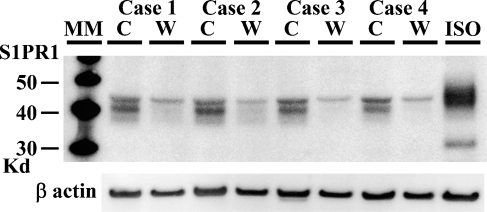
Figure 2.
A comparative study of S1PR1 expression between gray matter and white matter of cerebrum by Western blotting. All gray matter samples from four autopsy cases give strong signals at 40–45 kDa, whereas corresponding signals were faintly observed in white matter. Twenty μg of protein per lane was applied. Total proteins from an angiosarcoma cell line (ISO-HAS) were used as a positive control. Signals of β-actin appear with almost the same intensity between gray matter and white matter. C, cortex; W, white matter; ISO, ISO-HAS.

