
Figure 5. Sustained activation of p90RSK (Thr359/Ser363) in cortical and striatal neurons at 10 min reperfusion.
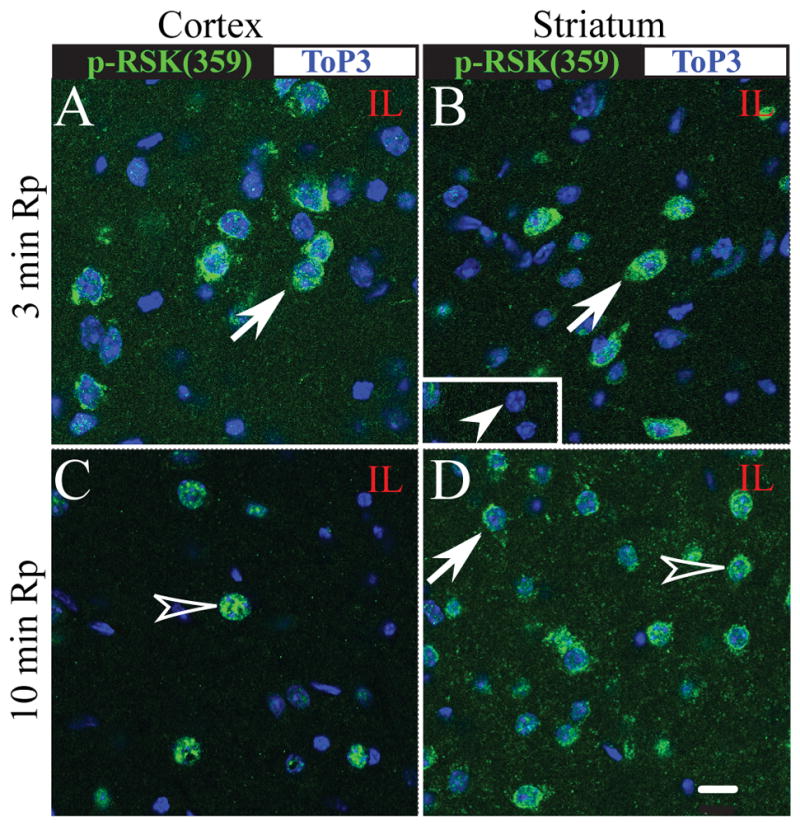
p-p90RSK (Ser359/363) expression (green) and Topro3 nuclear staining (blue) were shown in the ipsilateral cortex and striatum at 3 min Rp (A, B) or 10 min Rp (C, D). Arrow: increased immunoreactive signals of p-p90RSK. Open arrowhead: nuclear localization of p-p90RSK. Arrowhead in inset: an absence of p-p90RSK staining in the contralateral striatum. Identical digital imaging acquisition parameters were used in all images. Scale bar: 10 μm.