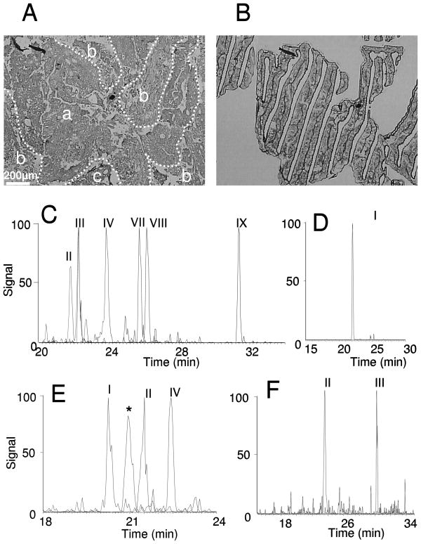
Figure 5. LC-MRM Detection of β-catenin, c-Src, c-Myc and PP2A in Laser Capture Microdissected Tumor Cells.
Optical microscope photographs (40× magnification) of colon adenocarcinoma tissue sections (A) stained with hematoxylin and dehydrated prior to LCM and extracted cancer cells on the LCM cap (B). In panel A, areas with colonic adenocarcinoma (a), tumor necrosis (b), and stroma (c) are indicated. Extracted ion chromatograms of detected peptides (see Table 1) from β-catenin (C), c-Myc (D), c-Src (E), and PP2A Catalytic subunit (F) are shown. The asterisks indicate interference in transitions from peptide, LLLNAENPR, from c-Src; however, the correct peak can be determined by comparison of fragment ion ratios in the composite tandem mass spectra.

