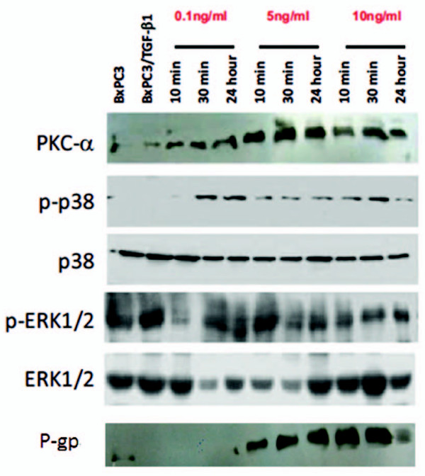
Figure 6.
The effects of TGF-β1 on expression levels of PKCα and p38 MAPK. BxPC3 cells were treated with 0.1, 1 and 10 ng/ml TGF-β1 for 10 min, 30 min and 24 h. Total cellular protein was extracted and subjected to western blotting analysis to detect expression of PKCα, phosphorylated-p38/total p38 MAPK and phosphorylated-ERK1/2/total ERK1/2. Bx represents BxPC3 cells and Bx/T represents the stably transfected BxPC3 cells with TGF-β1 plasmid.

