Figure 8.

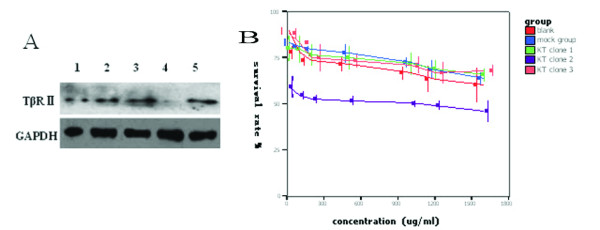
Role of TβRII siRNA in BxPC3 cells. (A) Western blotting analysis of TβRII (type II receptor or TGF-β1) protein levels. BxPC3 cells were grown and transfected with TβRII siRNA. After selection with G418, three clones were isolated and the cells from these clones underwent protein isolation. They were subjected to Western blotting analysis with anti-TβRII antibody. Lane 1, total pool of BxPC3 cells; lane 2, mock clone (transfected with empty plasmid, psilenser 2.1 U6); lane 3, knockdown (KD) clone 1; lane 4, KD clone 2; and lane 5, KD clone 3. (B) MTT assay. The transfected BxPC3 cells were grown and treated with gemcitabine at the indicated doses for 2 days. The cell viability was detected by using the MTT assay. The data show that inhibition of TβRII increases sensitivity of BxPC3 cells to gemcitabine. The IC50 value of clone 2 to gemcitabine was the lowest, indicating that clone 2 is more sensitive to gemcitabine than the other cells (P < 0.05).
