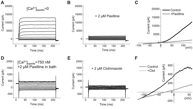
Figure 4. Functional expression of BK and IK1 channels in primary cells derived from glioblastoma multiforme (GBM).
(A) Representative recordings of macroscopic BK currents in primary cells cultured from surgical sample of glioblastoma multiforme. Currents were elicited by step pulses from −80 mV to +140 mV. (B) In the same cell shown in (A), the BK blocker paxilline (2 µM) potently inhibited macroscopic K+ currents. (C) Representative traces of K+ currents elicited in response to depolarization ramps from −80 mV to +140 mV in the absence and presence of paxilline. (D) Representative recordings of the whole-cell IK1 currents activated by step pulses from −120 to +60 mV. To isolate IK1 currents, [Ca2+]pipette was clamped at 750 nM and 2 µM paxilline was added into bath solution. (E) The specific IK1 inhibitor clotrimazole (2 µM) potently suppressed macroscopic K+ currents. (F) Representative traces of K+ currents elicited in response to depolarization ramps −120 mV to +60 mV in the absence and presence of clotrimazole. For additional experimental details, see legend to Fig. 2 and Results section.

