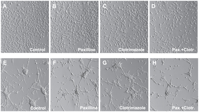
Figure 6. Representative micrographs of U251 (A-D) and U87 (E-H) cells grown in the presence of BK and IK1 blockers.
Cells were grown in the serum-free OptiMEM media supplemented with serum substitute B27. The BK blocker paxilline (10 µM) and the IK1 inhibitor clotrimazole (10 µM) were added as indicated. Images of the cells were captured ∼48 hrs after addition of channel blockers using Hoffman modulation contrast optics in Olympus IX71 microscope at 10×10 magnification.

