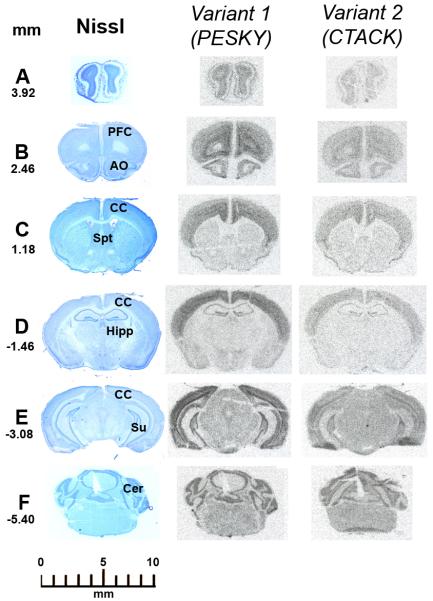
FIGURE 2.
Film autoradiographic images of the hybridization signal produced by radioactive riboprobes directed against the exon 1 of variant 1 or intron 1 of variant 2 of CCL27 in coronal serial consecutive sections of the murine brain. The left panel correspond to sections stained with Nissl for anatomical localization. The numbers represents the coronal levels in mm from Bregma. Section A is at the levels of the olfactory bulbs. Section B is at the level of the prefrontal cortex (PFC) and anterior olfactory nucleus (AO). Section C is at the level of the septum (Spt). Section D is at the level of the anterior hippocampus (Hipp). Section E is at the level of the posterior hippocampus and Subiculum (Su). Section F is at the level of the cerebellum (Cer). Specific signals for both variants are distributed trough the cerebral cortex (CC). For more detail see the text.

