Figure 1.
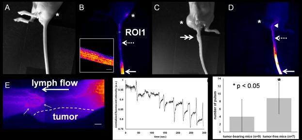
Abnormal lymphatic drainage pathways and tortuous, dilated collecting lymphatic vessels in tail-tumor-bearing mice. White light (A) and fluorescent (B) images in a tumor-free mouse showing fluorescent lymphatic capillaries (inset) near the injection site (arrow) and collecting lymphatic vessels (broken arrow) draining to the ischial LN (asterisk). Scale bar: 1 mm. White light (C) and fluorescent (D) images in a tail-tumor-bearing mouse depicting abnormal lymphatic passage to the abdominal region as denoted by the arrow head. Arrow: injection site. Double arrow: tumor. (E) Two fluorescent images stitched together in a tail-tumor-bearing mouse showing no uptake of fluorophore in the lymphatic capillaries in the skin above the tumor and tortuous and mispatterned collecting lymphatic vessels (small arrows) draining to the ischial LN. Flow is from right to left (distal to proximal). Scale bar: 1 mm. (F) Normalized fluorescent intensities as a function of time in the ROI1 selected on the fluorescent collecting lymphatic vessel shown in Figure 1B. (G) Collecting lymphatic vessels in tail tumor-bearing mice show significantly reduced number of contractions, as compared to those in normal mice. Error bars show the standard deviation.

