Figure 3.
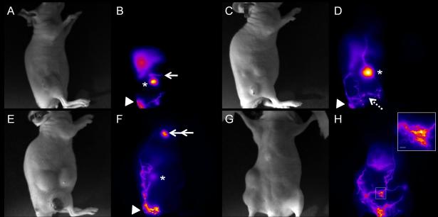
Abnormal lymphatic drainage and function and leaky lymphatic vessels in hindlimb-tumor-bearing mice. White light (A) and fluorescent (B) images in a mouse without palpable LN involvement at 2 weeks tumor p.i. show abnormal lymphatic drainage pathway (arrow) from the inguinal LN (asterisk). White light (C) and fluorescent (D) images in another mouse with palpable inguinal LN involvement at 2 weeks p.i. represent the enlarged fluorescent inguinal LN (asterisk) and leaky lymphatic vessels as indicated by bright spots (broken arrow). White light (E) and fluorescent (F) images in a mouse with the inguinal and axillary LN metastases showing dilated and tortuous fluorescent lymphatic vessels bypassing the primary tumor and the inguinal (Asterisk) and axillary LNs, and eventually draining to the brachial LN (double arrow). Arrow head: injection site. White light (G) and fluorescent (H) images of the ventral view of a tumor-bearing mouse with the massive inguinal and axillary LN metastases represent extensive ICG staining of lymphatic vessels, which are dilated and tortuous. The inset of Figure 3H shows a magnified fluorescent image of the rectangle in Figure 3H depicting diffused dye patterns. Scale bar: 1 mm.

