
Fig. 2.
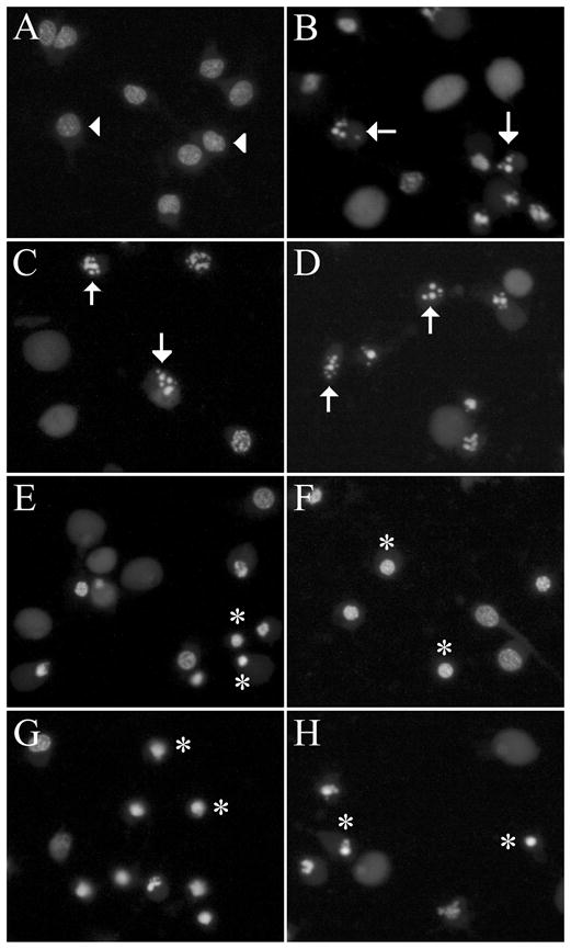
Cell death induced by Aβ, staurosporine, and Apoptosis Activator II is characterised by pyknotic nuclei. Cultures were treated with vehicle, 50 μM Aβ, 0.4 μM staurosporine (STS), 3 μM Apoptosis Activator II (AAII), 25 μM H2O2, 2.5 μM FeCl2/3, 200 nM A23187, or 2.5 mM 3-NP for 24 h. Representative images show nuclear changes visualised with membrane-permeable nucleic acid stain SYTO 11. (a) Vehicle-treated control neurones show normal morphology (arrowheads). (b) Aβ, (c) staurosporine, and (d) Apoptosis Activator II treatment induce pyknotic nuclei in neurones (arrows), while (e) H2O2, (f) FeCl2/3, (g) A23187, and (h) 3-NP induce condensed nuclei (asterisks).