
Figure 2. Atoh1 misexpression generates supernumerary Myo7a+ cells bearing stereociliary bundles.
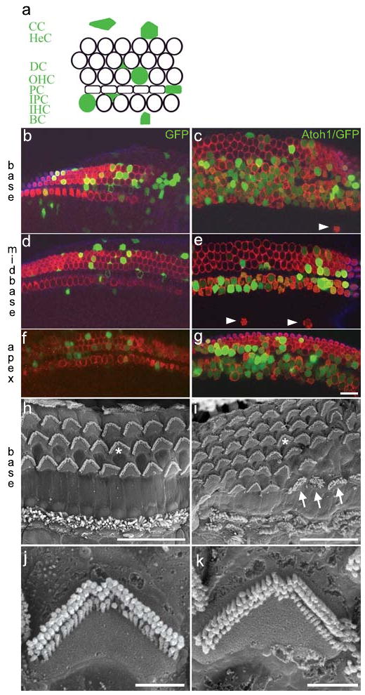
a, schematic of cell types in the organ of Corti transfected with Atoh1/enhanced GFP (filled green): CC, Claudius' cells; HeC, Hensens' cells; DC, Deiters' cells; OHC, outer hair cell; PC, pillar cell; IPC, inner phalangial cell; IHC, inner hair cell; BC, border cell. b-g, laser confocal micrographs of E18.5 GFP-transfected (b,d,f) and Atoh1/GFP-transfected (c,e,g) organs of Corti immunostained for Myo7a (red). All of the Atoh1/GFP+ cells in panels c,e, and g are Myo7a+. h,i, scanning electron micrographs of postnatal day 35, untransfected (h) and Atoh1/GFP-transfected (i) organs of Corti. Asterisks in panels h,i indicate stereociliary bundles imaged at higher magnification in panels j,k, respectively. The arrows indicate 3 cells with atypical bundles. Scale bars: b-i, 20μm; j,k, 2 μm.