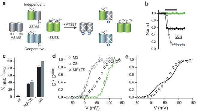
Figure 3.
Cooperative gating in Hv1 dimers made of MS and ZS subunits. (a) Assembly of MTSET-sensitive (MS) and Zinc-sensitive (ZS) subunits in homo-and heterodimers. White X indicates blocked MS subunit after MTSET treatment. (b) Example of changes in normalized proton currents from membrane patches of oocytes expressing Hv1 MS (gray diamonds), Hv1 ZS (green squares), or co-expressing Hv1 MS with ZS (black circles). 25 μM Zn2+ was maintained in the extracellular solution throughout the recordings. pHi = pHo = 6.0. Black bar indicates duration of exposure to 1 mM intracellular MTSET. The RNA ratio for the co-expression of MS and ZS subunits was 3:1. (c) Quantification of inhibition produced by MTSET for the indicated conditions (black bars). From the extent of proton current inhibition the percentage of MS subunits was calculated (gray bars), as explained in the text. (d) Voltage dependence of proton conductance of Hv1 MS and Hv1 ZS, compared to the voltage dependence of the co-injection of Hv1 MS + Hv1 ZS. See Table 1 for Boltzmann fits. Each point is the average of 4–6 measurements ± s.e.m. Recording conditions and RNA ratio were the same as for panels b and c. (e) Comparison between voltage dependence of proton channels produced by co-expression of MS and ZS subunits (open circles), and voltage dependence expected in case of independent opening of the two pores (continuous line, see text).

