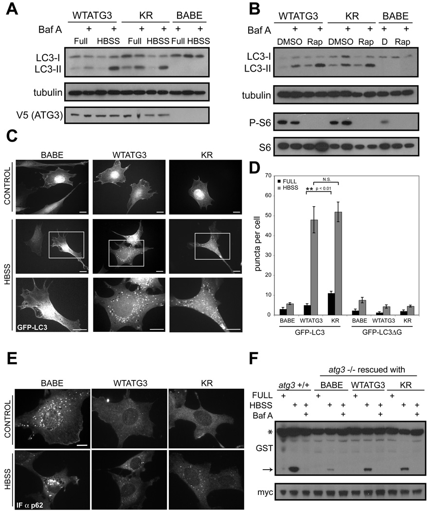
Figure 4. Starvation-induced autophagy remains intact upon disrupting ATG12 conjugation to ATG3.
Stable pools of atg3−/− fibroblasts expressing an empty vector (BABE), wild type mouse ATG3 (WTATG3) or mutant ATG3 unable to be conjugated by ATG12 (KR) were used for experiments as indicated. (A and B) Cells were grown in complete media, starved in Hank’s buffered salt solution (HBSS) for 4h, or treated with 10nM rapamycin (B) for 6h. Cells were lysed and immunoblotted with indicated antibodies. Phosphorylated ribosomal S6 (P-S6) was used to verify rapamycin-mediated mTORC1 inhibition. When indicated, bafilomycin A (BafA, 10nM) was added to cells 1h prior to lysis. (C) Indicated cells types expressing GFP-LC3 were grown in complete media (control) or HBSS-starved for 4h; boxed areas from center panels are enlarged below. Bars, 25µm. (D) Quantification of punctate GFP-LC3 or GFP-LC3ΔG (mean +/−SEM puncta per cell). (E) Indicated cell types grown in complete media (control) or HBSS-starved for 4h, and then fixed and immunostained with α-p62 antibody. Bar, 25µm. (F) Indicated cell types were transfected with a GST-BHMT fusion construct, HBSS-starved for 6h, lysed and immunoblotted with α-GST. Asterisk (*) indicates full-length GST-BHMT and arrow indicates cleaved BHMT produced in autolysosomes. When indicated, bafilomycin A (BafA, 10nM) was used to inhibit lysosomal function. α-Myc was used to detect GFP-myc (expressed from an IRES sequence) to control for transfection efficiency (Dennis and Mercer, 2009). See also Figure S3.

