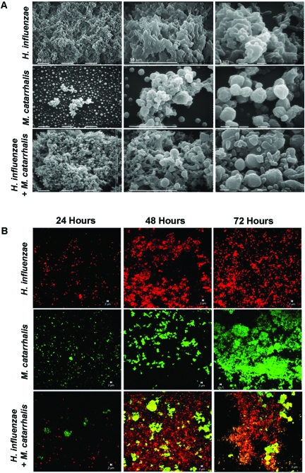
FIG 1 .
H. influenzae and M. catarrhalis form polymicrobial biofilms in vitro. Stationary biofilms were established in chamber slides for visualization of bacteria by SEM and confocal laser scanning microscopy (CLSM). (A) Samples of H. influenzae and M. catarrhalis single-species or polymicrobial biofilms were taken at 48 h and prepared for SEM. Images shown are at three different levels of magnification. (B) CLSM was performed on 24-, 48-, and 72-h biofilms following staining of H. influenzae (red) and M. catarrhalis (green).

