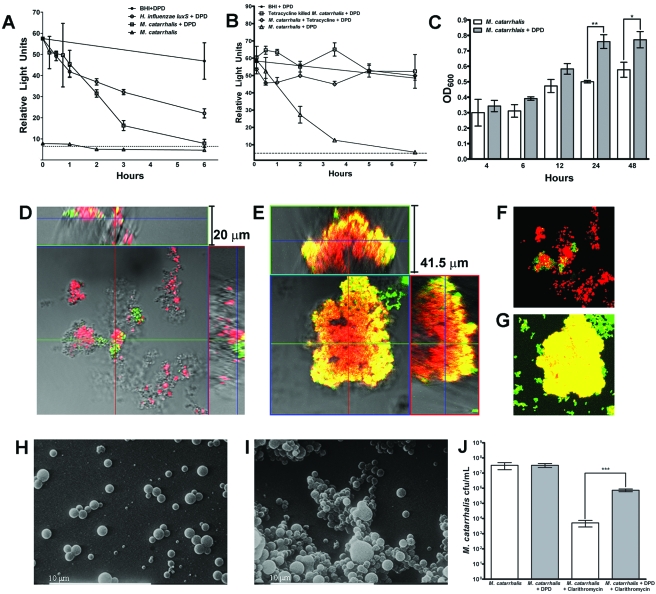
FIG 4 .
AI-2 promotes M. catarrhalis biofilm development and antibiotic resistance. (A) M. catarrhalis was cultured in BHI medium or BHI medium supplemented with 0.2 µM synthetic AI-2 (DPD) to determine AI-2 production and depletion, as measured by Vibrio harveyi bioluminescence. H. influenzae luxS was cultured in sBHI medium supplemented with DPD to measure depletion. An uninoculated control of BHI medium with DPD shows the minimal degradation of the AI-2 signal during 6 h of incubation at 37°C. (B) Depletion of DPD by M. catarrhalis biofilms were established for 24 h following incubation with 10 µg/ml tetracycline was measured by bioluminescence over a period of 7 h. (C) M. catarrhalis biofilms were established in the presence or absense of DPD and stained with crystal violet for determination of biofilm biomass at 4, 6, 12, 24, and 48 h. Data represent the mean results from three combined experiments, with three replicate wells per experiment. Error bars represent SEM. (D and E) M. catarrhalis biofilms were established for 24 h in the presence (E) or absence (D) of DPD and stained with a viability kit for CLSM visualization of surface coverage and biofilm thickness. (F and G) Z-series images from panels D and E were compressed to show total viable and nonviable staining of biofilms established in the presence (G) or absence (F) of DPD. (H and I) SEM images of 24-h M. catarrhalis biofilms established with (I) or without (H) DPD. (J) M. catarrhalis biofilms were established for 4 h in the presence or absence of DPD and then treated with 6 µg/ml clarithromycin for 24 h and plated for enumeration of viable bacteria. Data represent the means from three replicates ± SEM. *, P < 0.05; **, P < 0.01; ***, P < 0.001.

