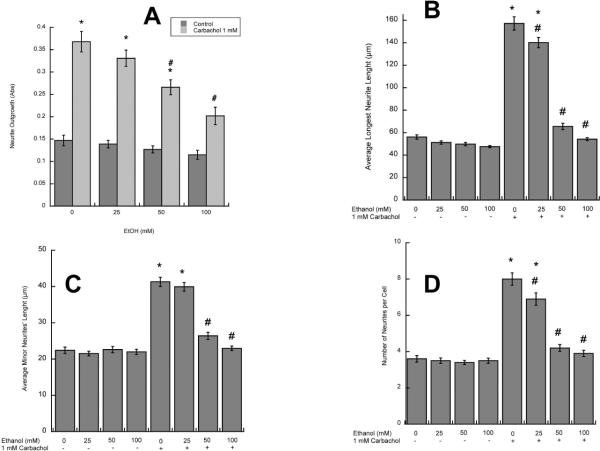
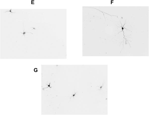
Figure 1. Effect of ethanol on carbachol-treated astrocyte-induced hippocampal neuron neurite outgrowth.
A: Hippocampal neurons were incubated in the presence of astrocytes previously treated with 1 mM carbachol and/or 25, 50, or 100 mM ethanol. (A): Neurite proteins were quantified spectrophotometrically as described in Methods (n=6). (B, C, D): Morphometric analysis was carried out in hippocampal neurons immunostained with a neuron-specific βIII-tubulin antibody. Pictures were taken with a digital camera attached to a fluorescence microscope. Quantifications of the length of the longest neurite (A) and the length of minor neurites (B) were carried out using the software MetaMorph. C: number of processes per cell. The results (mean +/− S.E.) derive from the measurements of 60–80 cells per treatment. Representative fields of hippocampal neurons co-cultured with control (D), carbachol-treated (E) or carbachol- and 50 mM ethanol-treated (F) astrocytes are also shown.
*: p<0.05 vs control; #: p<0.05 vs carbachol by the Dunnett post-hoc test.


