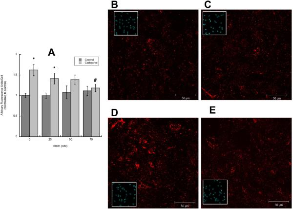
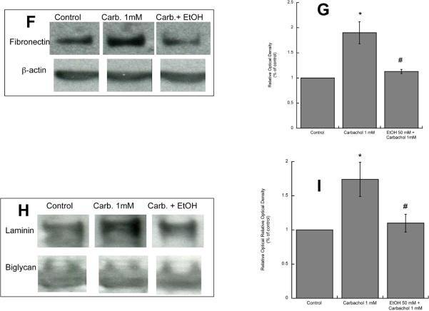
Figure 3. Effect of ethanol on carbachol-induced increase in extracellular laminin.
A: Astrocytes were stimulated for 24 h with 1 mM carbachol alone or in the presence of 25, 50, or 75 mM ethanol. After immunolabeling with a laminin-1 antibody, cells were analyzed by confocal microscopy. Average laminin immunofluorescence per cell was quantified as described in “Methods”. *: p<0.05, vs. control; #: p<0.05 vs. carbachol by the Dunnett post hoc test (n=6). Representative fields of astrocytes treated with serum-free medium (Control; B); 50 mM ethanol (C); 1 mM carbachol (D); and 1 mM carbachol in the presence of 50 mM ethanol (E) are shown. (F, G,): Western blot analysis was carried out in the cell lysate of astrocytes treated for 24 h with 1 mM carbachol in the presence or absence of 50 mM ethanol. After protein transfer, membranes were labeled with a laminin (F, upper blot) or β-actin (F, lower blot) antibody. (G): Densitometric analysis of cellular levels of laminin normalized to β-actin is shown. (H, I): Proteins present in astrocyte-conditioned medium were separated by electrophoresis, transferred to PVDF membranes, and labeled with a laminin (H, upper blot) of byglican (H, lower blot) antibody and detected by Western blot. (I): Densitometric analysis of laminin in astrocyte-conditioned medium normalized to byglican is shown. *, p<0.05 vs. control; #, p<0.05 vs. carbachol (n=3).


