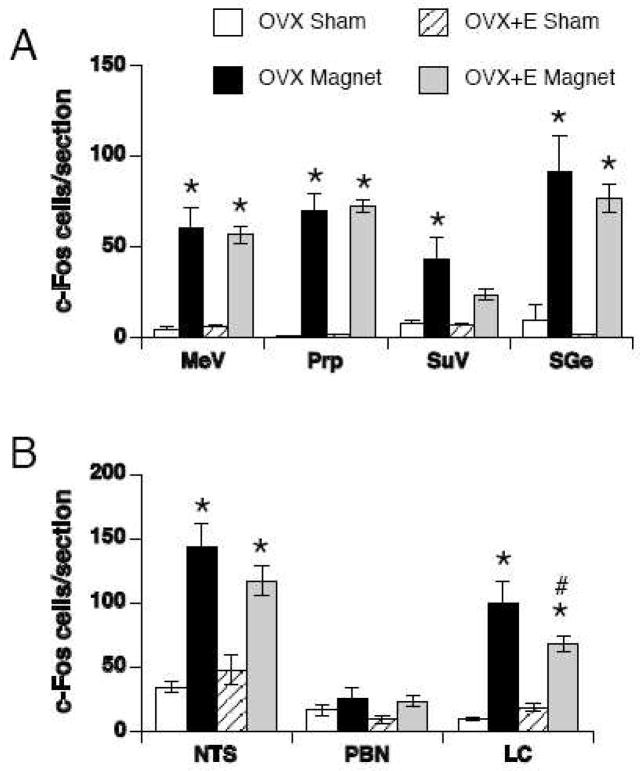
Figure 4.
Quantification of c-Fos positive cells in sham-exposed (white and striped bars) or magnet-exposed (black and gray bars) rats in vestibular (A) and visceral relays (B). The bilateral counts (mean ± SEM) through each section are shown. MeV = medial vestibular nucleus, Prp = prepositus nucleus, SuV = superior vestibular nucleus, SGe = supragenualis nucleus, NTS = nucleus of the solitary tract, PBN = parabrachial nucleus, LC = locus coeruleus. * p < 0.05 within group v. sham-exposed rats; # p < 0.05 within magnet-exposed rats, OVX v. OVX+E.

