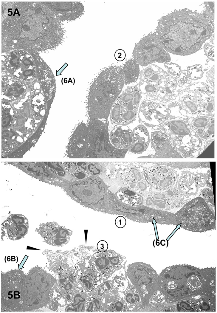
Figure 5.
Results of the PMN response to chlamydiae-infected conjunctival epithelial cells. (A) PMNs apparently in the act of “pushing” the infected epithelial cells off the mucosal lining, causing a breech in the barrier with (B) subsequent release of intact, infected epithelial cells, damaged epithelial cells, chlamydiae (arrowheads) and PMNs. One can discern a progression in events (as denoted by the circled numbers) in which 1) PMNs accumulate under an intact infected epithelium; 2) the epithelial cell layer begins to lose its integrity; and 3) the epithelium has been breached, releasing PMNs onto the surface. Arrows denote examples of chlamydiae-infected superficial epithelial cells, which are more clearly evident in enlarged images in Figures 6A–C. Magnification: × 3,760.

