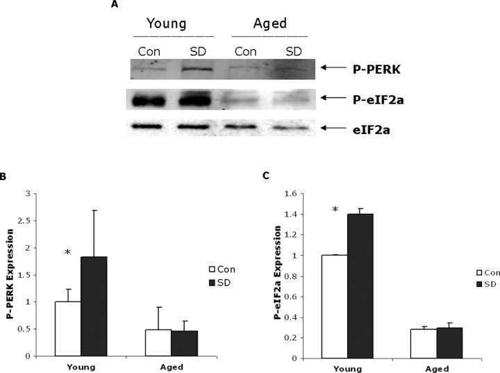
Figure 4.
A, PERK and eIF2α phosphorylation in young and aged cerebral mouse cortex. Western blot showing P-PERK and phospho-eIF2α expression in young and aged samples (20 μg of protein loaded per well) with and without sleep deprivation (SD). eIF2α levels shown below are used as loading controls (Con). B, Densitometric quantification of P-PERK signal directly from chemiluminescence in young and aged sleep-deprived and control samples; mean values with standard deviation shown (n = 6, *p = 0.01). C, The level of phosphorylated eIF2α is decreased in aged mouse cerebral cortex. Densitometric quantification of P-eIF2α signal directly from chemiluminescence in young and aged sleep-deprived and control samples; mean values with standard deviation are shown (n = 8; *p < 0.0001).

