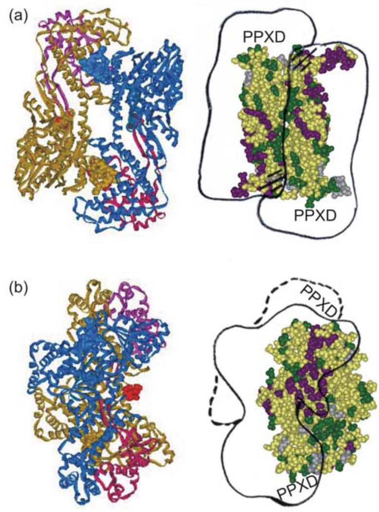
Figure 11.
Simultaneous binding of precursor galactose-binding protein and SecA. Panel (a) left side, structure of the SecA dimer oriented so that the extreme C-termini, which are not resolved, and the PPXD (magenta and pink) are on the lower surface. Right side, an outline of the SecA dimer is docked across SecB as described in the text. The hatched lines indicate the binding sites on SecA that interact with the flexible C-terminal regions of SecB (shown in structure as helical). Panel (b) structures shown are related to those in panel (a) by a 90° rotation about the vertical axis to the left. The PDB code for SecA is 1M74.

