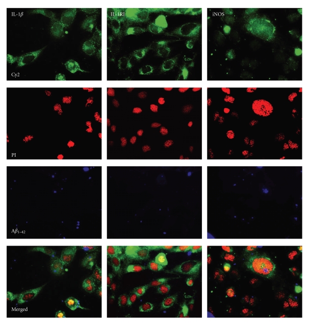
Figure 6.
Uptake of Aβ 1–42 and expression of cellular markers in human microglial cells. The micrographs show human CHME3 microglial cells after incubation with biotinylated Aβ 1–42, after which they were fixed and stained with antibodies against interleukin-1β (IL-1β), IL-1 receptor type I (IL-1RI) and inducible nitric oxide synthase (iNOS), and subsequent incubation with Cy2-conjugated secondary antibodies. Cell nuclei were stained with propidium iodide (PI) and the biotinylated Aβ 1–42 was visualized with AMCA-conjugated streptavidin. All micrographs are in 20× magnification.

