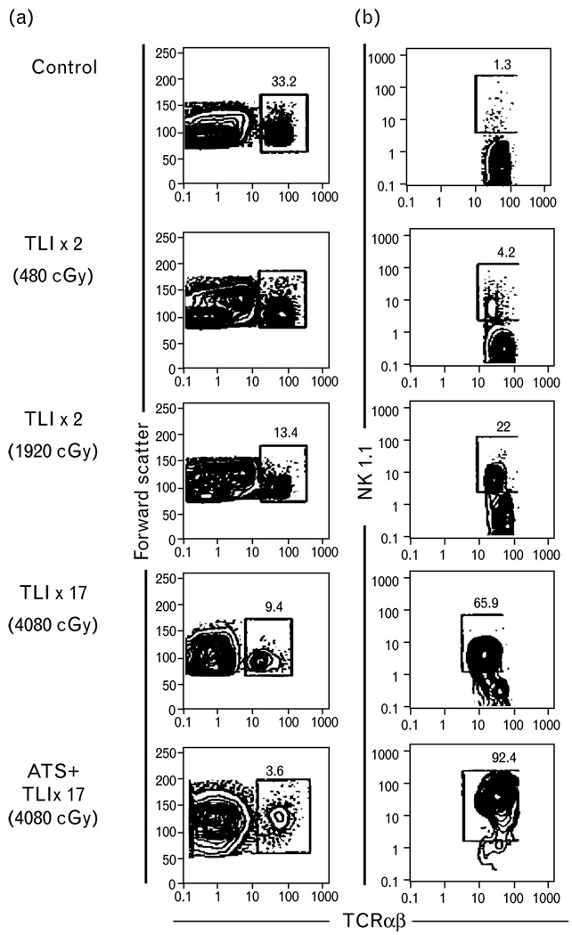
Figure 1. Immunofluorescent staining of C57BL/6 spleen cells.
(a) Immunofluorescent staining of C57BL/6 spleen cells after 2, 8, or 17 doses of TLI or TLI and ATS. Flow cytometric analysis shows light scatter versus TCRαβ or NK1.1 versus TCR αβmarkers. (b) Staining of BALB/c spleen cells for DX5 and TCRαβ markers. Boxes show percentages of TCRαβ cells, and percentage of NK1.1+ or DX5+ cells amongst gated TCRαβ+ cells [6]. ATS, antithymocyteserum; TLI, total lymphoidirradiation.

