Figure 1.
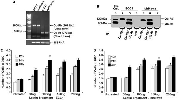
Expression of leptin receptor and effect of leptin on the proliferation of endometrial cancer cells. (A) Total RNA was extracted from ECC1 and Ishikawa cells and analyzed by RT-PCR using specific primers for long isoform (Ob-Rb) and short isoform (Ob-Rt) leptin receptors. A primer set for 18S RNA was used as a control. (B) Total protein was isolated from ECC1 and Ishikawa cells and equal amounts of proteins were subjected to immunoprecipitation (IP) using specific antibodies for long and short forms of the leptin receptor. Immunoprecipitation with IgG was included as a negative control and 25 μg of COLO 320DM cell lysate (Santa Cruz Biotechnology) were included as a positive control. Immunoprecipitates were resolved by SDS-PAGE and subjected to immunoblot analysis using a mouse monoclonal antibody against both long and short forms of leptin receptor. Ob-Rb and Ob-Rt were found to be present in ECC1 and Ishikawa cells. (C) ECC1 cells and (D) Ishikawa cells were serum starved for 16 h followed by treatment with 50–200 ng/ml leptin for 12, 24 and 48 h. XTT assays were then performed as described in ‘Materials and methods’. Leptin treatment increased proliferation of ECC1 and Ishikawa cells in a time- and dose-dependent manner. *P < 0.01, for different times and doses compared with respective untreated cells. The data represent mean values±s.e.m. and are the results of three independent experiments performed in triplicates.

