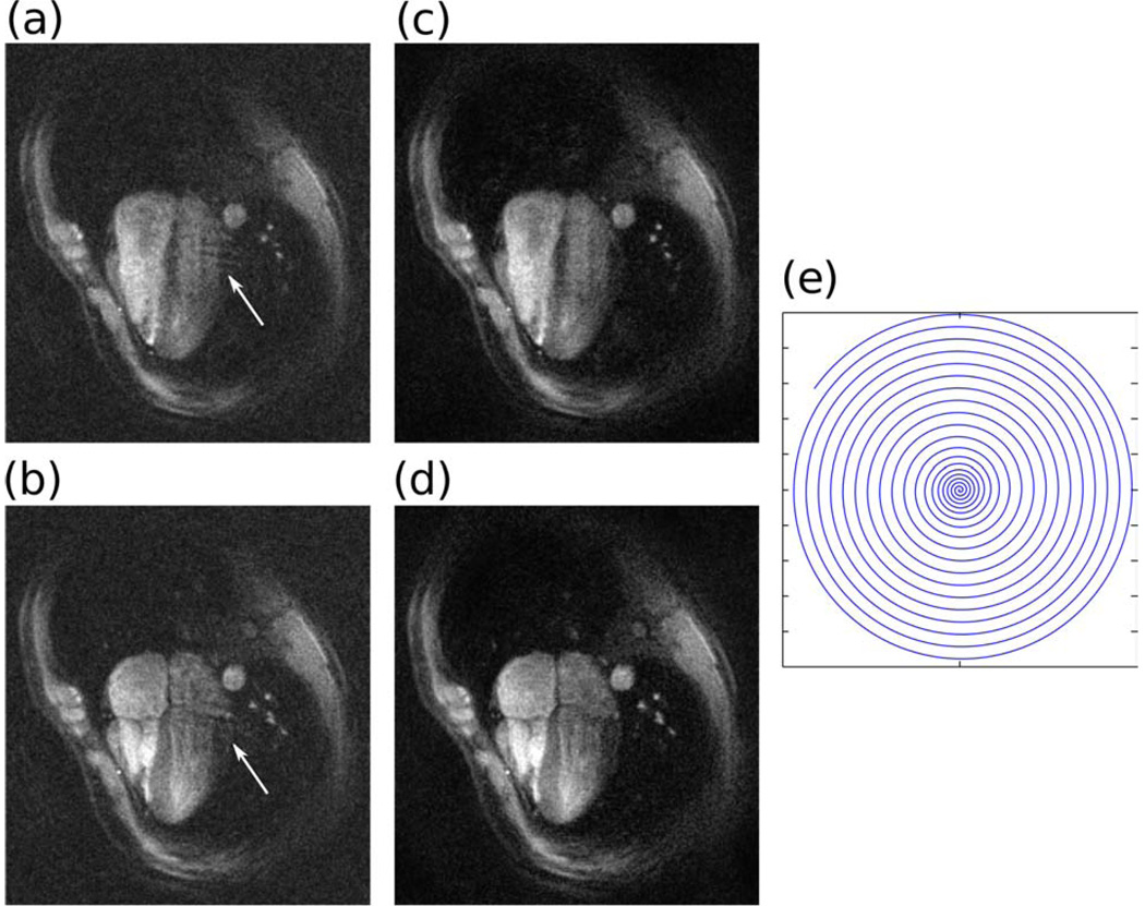
Figure 7.
Dynamic cardiac imaging with dual density spirals with 3-fold acceleration and 4 channels. Two phases of a four chamber view of the heart. (a)–(b) Sum-of-squares of gridding reconstruction exhibits coherent (arrows) and incoherent (noise-like) aliasing artifacts. (c)–(d) Both the coherent and incoherent artifacts are removed by SPIRiT. (e) One out of the three spiral interleaves.

