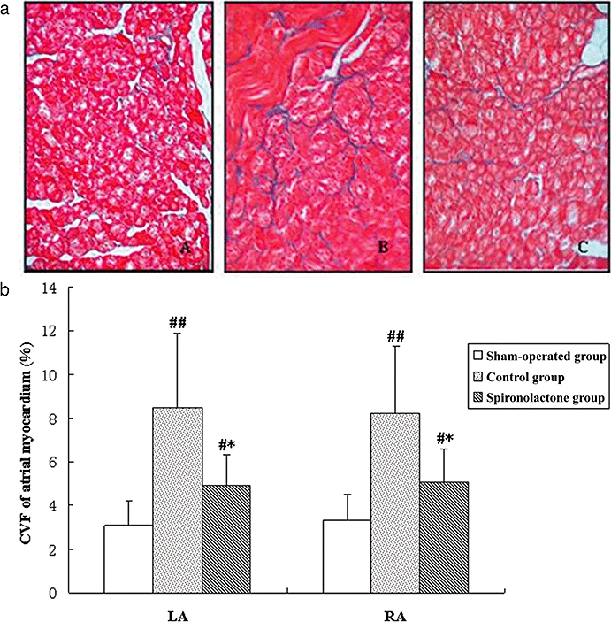
Figure 2.

(a) Representative Masson's trichrome staining of atrial myocardium. Red areas represent myocytes and blue areas represent collagen. A: the sham-operated group; B: the control group; C: the spironolactone group. In sham-operated group (A) almost no interstitial fibrosis was present. Extensive fibrosis was especially visible around myocytes of paced atria (B), but attenuated in the spironolactone group (C) (Masson trichrome staining, original magnification ×400). (b) Collagen volume fractions (CVF) of atrial myocardium. After rapid atrial pacing, CVF in control group increased significantly compared with sham-operated group. Spironolactone treatment decreased CVF of LA and RA compared with the control group. #P < 0.05, ##P < 0.01, compared with the sham-operated group; *P < 0.05, compared with the control group. LA, left atrium; RA, right atrium.
