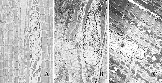
Figure 3.

Typical ultrastructural changes by transmission electron microscopy. A: the sham-operated group; B: the control group; C: the spironolactone group. Atrial myocytes from sham-operated dogs (A) had regular sarcomere organization, uniformly sized mitochondria and a normal nucleus. Samples of atrial tissue taken from control group (B) showed abnormal ultrastructure: severe disintegration of myofilaments and loss of banding pattern and integrity of contractile elements, mitochondrial swelling with a decrease in the density and organization of the cristae, karyopyknosis with chromatin margination to nuclear membrane indicating cell apoptosis. These chronic pacing-induced ultrastructural changes were markedly attenuated by treatment with spironolactone (C) (magnification: A: ×6000; B and C: ×5000).
