
Figure 7.
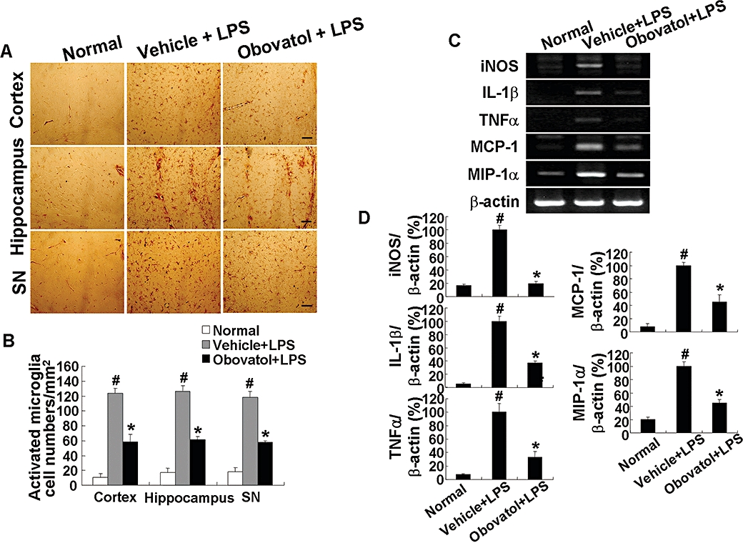
Obovatol suppressed microglial activation in a mouse neuroinflammation model. C57BL/6 mice were injected i.p. with vehicle (saline containing 0.5% DMSO and 5% propylene glycol) or obovatol (diluted in saline containing 5% propylene glycol) once daily at 10 mg·kg−1 for 4 days. At 24 h after the first injection of vehicle or obovatol, mice were injected i.p.with 5 mg·kg−1 LPS. Mice were anesthetized with diethyl ether and transcardially perfused with ice-cold saline 72 h after the LPS injection. Brains were removed and sections were stained with IB4 (a marker for microglia). IB4-positive cells were observed in cortex, hippocampus, and substantia nigra (SN) region of vehicle+LPS- or obovatol+LPS-injected mouse brains (A). Scale bar, 50 µm. The number of IB4-stained cell per mm2 was counted (B). The expression levels of proinflammatory genes were determined by RT-PCR at 6 h after the LPS injection (C). Levels of iNOS, IL-1β, TNFα, MCP-1 and MIP-1α were normalized to β-actin levels and expressed as a relative change in comparison with the LPS treatment, which was set to 100% (D). The data were expressed as the mean ± SD (n= 3 per experimental group). #P < 0.01 versus normal animals; *P < 0.01 versus vehicle+LPS-injected animals. DMSO, dimethyl sulfoxide; LPS, lipopolysaccharide; RT-PCR, reverse transcription-polymerase chain reaction; iNOS, inducible nitric oxide synthase; IL-1, interleukin 1; TNF, tumour necrosis factor; MCP, monocyte chemotactic protein; MIP, macrophage inflammatory protein.