
Figure 10.
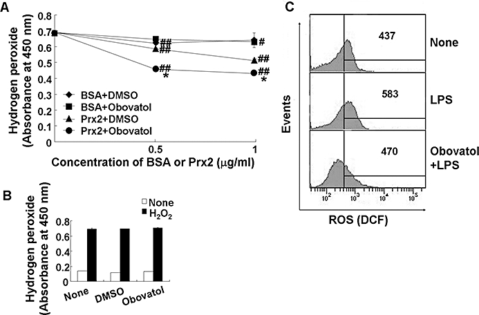
Obovatol enhanced the peroxidase activity of Prx2 and suppressed intracellular ROS production. Ferrous oxidation assay was carried out in 100 µL of reaction mixture containing DTT (2 mM), H2O2 (50 µM), obovatol (10 µM) and recombinant Prx2 protein. After incubation of the mixture for 10 min, the remaining H2O2 was measured based on the color changes after adding Fe(NH4)SO4 and potassium thiocyanate. DMSO and BSA were used as a vehicle control for obovatol and a negative control for Prx2 respectively. The data were expressed as the mean ± SD (n= 3). #P < 0.05, ##P < 0.01 versus H2O2 only; *P < 0.01 versus BSA+DMSO or Prx2+DMSO (A). The direct H2O2-scavenging effect of obovatol was also evaluated by ferrous oxidation assay in the absence of recombinant Prx2 protein (B). BV-2 microglia cells were treated with 100 ng·mL−1 LPS for 30 min in the absence or presence of obovatol (10 µM), followed by 30 min incubation with 10 µM H2DCF-DA. Fluorescence was measured by flow cytometer. The results were expressed as the mean fluorescence intensity (C). DMSO, dimethyl sulfoxide; Prx2, peroxiredoxin 2; ROS, reactive oxygen species; DTT, dithiothreitol; BSA, bovine serum albumin.