
Figure 11.
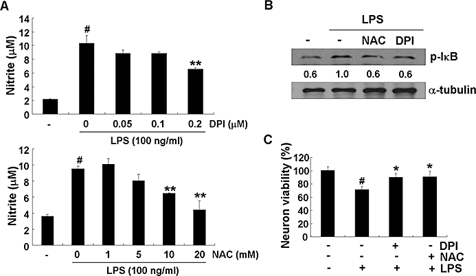
NADPH oxidase mediated LPS-induced microglial activation. BV-2 microglia cells were treated with DPI or NAC for 30 min before the addition of LPS (100 ng·mL−1). After 24 h, the nitrite in the media was measured by Griess reaction (A). After 30 min activation with LPS (100 ng·mL−1), the cells were harvested and analysed by immunoblot for phospho-IκB. Levels of phospho-IκB were normalized to α-tubulin levels and expressed as a fold decrease compared with the LPS treatment (B). Primary microglia were stimulated with LPS (100 ng·mL−1) in the absence or presence of DPI or NAC for 6 h, culture media were replaced with fresh media and further incubated for 24 h. The microglia-conditioned media were collected and transferred to primary neurons followed by additional 24 h incubation. At the end of incubation, cell viability was determined by MTT. Results were expressed as a percentage of control (mean ± SD) (C). The data were expressed as the mean ± SD (n= 3). #P < 0.01 versus untreated control; *P < 0.05, **P < 0.01 versus LPS only. LPS, lipopolysaccharide; DPI, diphenyliodonium; NAC, N-acetyl cysteine.