
Figure 6.
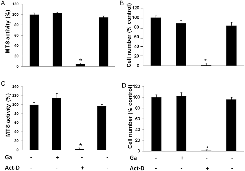
Effect of Ga on osteoblastic viability and proliferation. Murine MC3T3-E1 osteoblastic cells (A, B) and murine primary calvaria-derived osteoblasts (C, D) were cultured as described in the Materials and Methods. After overnight incubation, cells were treated with 100 µM Ga or 5 µg·mL−1 Act-D (+) or their respective vehicles (−) for 48 h. Osteoblastic viability was evaluated by MTS activity. Results are expressed in relative MTS activity compared with untreated cells (*P < 0.05 significantly different from untreated cells) (A, C). Osteoblastic proliferation was quantified by scoring cells manually after Trypan blue staining. Results are expressed as relative cell number as compared with untreated cells (B, D). *P < 0.05 significantly different from corresponding untreated cells. Ga, gallium; MTS, methyl tetrazolium salt.