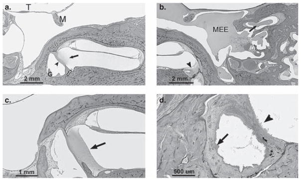
Figure 1.
(a) Section of temporal bone showing inflammation extending into the scala tympani (arrow) via an inflamed round window membrane (arrowhead). There is granulation tissue (G) in the round window niche and inflammatory cells in the cochlear aqueduct area (open arrow). (b) Section showing effusion in the middle ear cavity (MEE) and within the mastoid air cells (arrow). Effusion is also seen in the round window niche and in the perilymphatic space of the cochlea (arrowhead) across the round window membrane. (c) Section showing serofibrinous precipitate (arrow) in the scala tympani (basal turn) adjacent to the round window membrane. The round window membrane appears partly obliterated by the precipitate. (d) Section showing marked thickening and infiltration of the round window niche mucosa by inflammatory cells (arrow). Inflammatory cells are seen lining the perilymphatic space (arrowhead) of the inner ear adjacent to the round window membrane. Staining with H&E. T, tympanic membrane; M, malleus.

