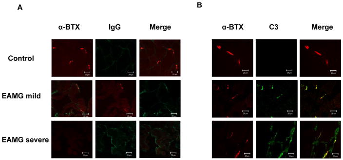
Figure 4.
Depostion of IgG (left) and C3 (right) at the postsynaptic endplate. Endplates from control animals show no significant deposition of IgG or C3 (top rows). Mice with mild EAMG show strong staining for IgG and C3 which co-localizes to endplate regions as defined by staining with tetramethylrhodamine-conjugated α-BTX (middle rows). Mice with severe EAMG show staining for IgG and C3 which appears to target the nonjunctional muscle fiber, failing to co-localize to the endplate region (bottom rows).

