Figure 3.
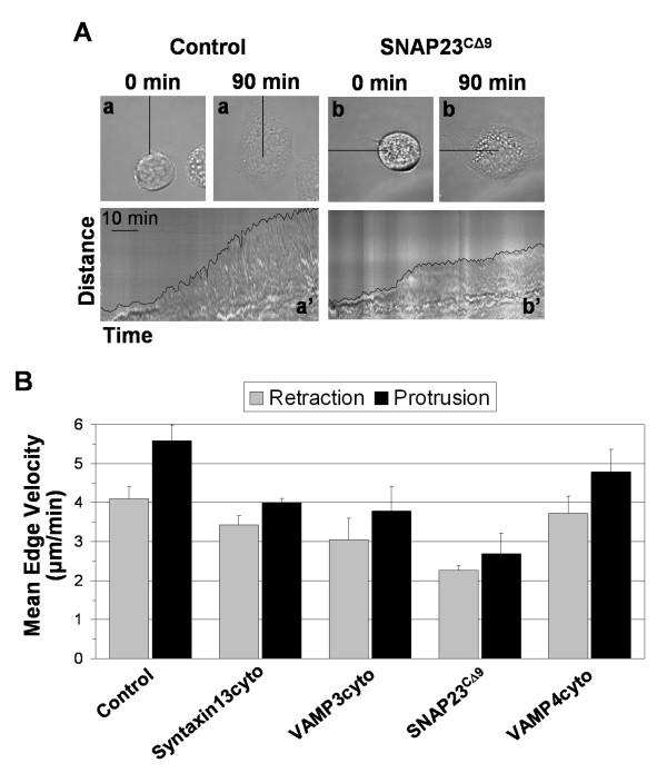
Inhibition of SNAREs reduces the rate of lamellipodium protrusion. Transfected CHO-K1 cells were plated in serum-free media on fibronetin for 90 min and monitored in real-time. (A) Representative control (a) and SNAP23CΔ9 (b) cells are shown at initial acquisition (0 min) and after 90 min (90 min) in the top panels. Kymographs (a' and b') in lower panels represent progression of the cell edge along the lines indicated with the cell edge highlighted. (B) The protrusion and retraction rate of the cell edge was determined from kymographs using ImageJ software. Data is presented as the mean velocity of immediate edge movement from at least two cell edges from at least 3 cells (when possible the most protrusive regions of the cell were measured).

