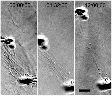
Figure 2. DIC image showing pipette drift stretching fibrils to over 100% initial length during enzymatic degradation.
Experimental time series images show the micropipettes drifting 30–40 µm apart, without fracturing or significantly thinning attached collagen fibrils, indicating either additional collagen deposition or structural changes within the fibrils. Note, in the last frame, stretched fibrils remain visible in the microbioreactor while peripheral fibrils have degraded. Experiments with large pipette drift were excluded from analysis. Bar = 10 µm.

