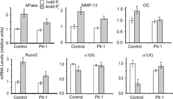
Figure 4.
The effect of suppression of Pit-1 expression on extracellular Pi-mediated stimulation of terminal differentiation markers, including APase, MMP-13, osteocalcin (OC), runx2, type X collagen (α1(X)), and type II collagen (α1(II)). Pit-1 expression was suppressed by specific siRNA (Pit-1). Control cells were transfected with a control siRNA (Control). After 2 days of transfection, cells were cultured in the presence of 1mM or 4mM Pi. The levels of hypertrophic and terminal differentiation marker mRNAs were determined by real-time PCR and SYBR Green and normalized to the 18S RNA levels. Data are means of triplicate PCRs using RNA from three different cultures; error bars represent standard deviations (ap < 0.01 vs. 1mM Pi-treated/control siRNA-transfected-cells; bp < 0.01 vs. 4mM Pi-treated/control siRNA-transfected cells)

