
Figure 1.
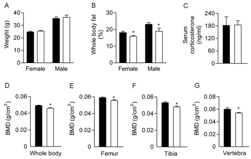
Phenotypic analysis of 7-week-old Col3.6-HSD2 (TG) and wild type (WT) littermates. A) Body weight. B) Percent body fat analyzed by DEXA. C) Serum corticosterone levels in female mice. D–G) BMD of whole body, femurs, tibiae and vertebrae in female mice assessed by DEXA. Each value is the mean ± SD for 5–10 mice. Black bars, WT; White bars, TG. *Different than WT, p<0.05.