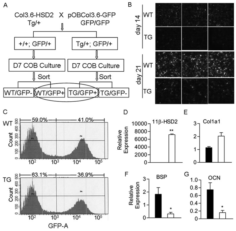
Figure 4.

Gene expression in primary osteoblast cultures carrying a pOBCol3.6-GFP transgene. A) Heterozygous Col3.6-HSD2 mice were crossed with homozygous pOBCol3.6-GFP mice to yield mice that were all heterozygous for the pOBCol3.6-GFP transgene and either wild type (WT) or transgenic (TG) for the Col3.6-HSD2 transgene. Primary calvarial osteoblasts (COB) were prepared from the mice and cultured 7 days. Cells were sorted based on GFP and the WT/GFP+ and TG/GFP+ populations were used for gene expression studies. B) GFP fluorescence in day 14 and 21 cultures of unsorted COB cells prepared from WT and TG female mice. C) FACS analysis of GFP expression in sorted WT/GFP+ and TG/GFP+ day 7 cultures. D–G) Expression of the 11β-HSD2 transgene and osteoblast genes in sorted WT/GFP+ and TG/GFP+ cells. Black bars, WT/GFP+ cells; White bars, TG/GFP+ cells. Each value is the mean ± SD for 3 samples and each experiment has been performed at least twice with similar results. *Different than WT/GFP+, *p<0.05, **p<0.01.
