
Figure 4.
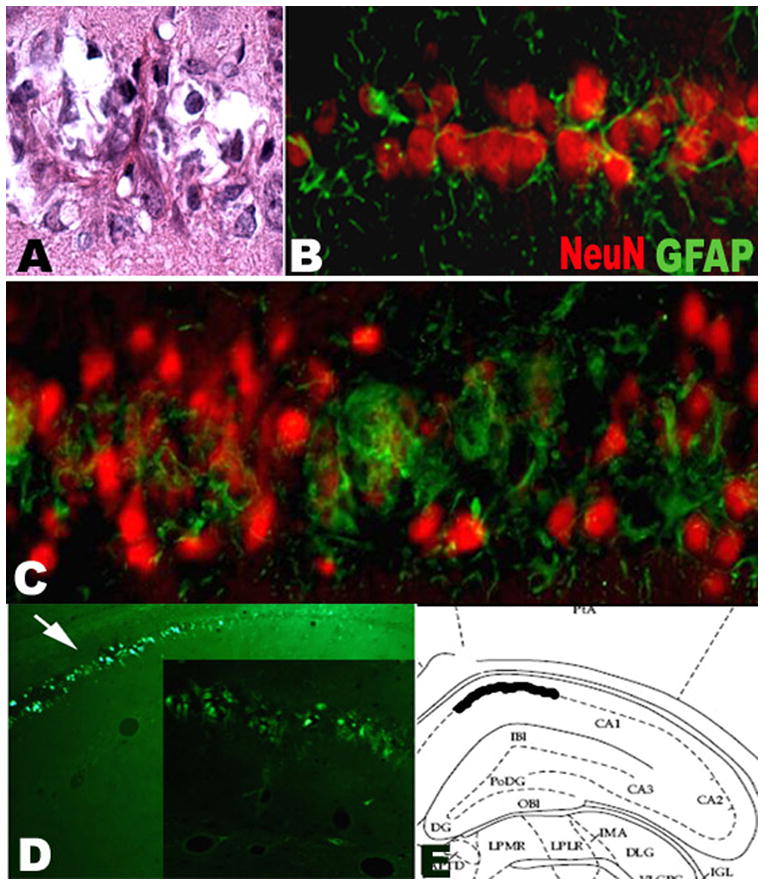
A: H&E showing damage of CA-1 in a rat from LPS+LiPC group. A small cell with acidophilic cytoplasm is surrounded by a fibrillar structure. B: Double immunostaining NeuN-GFAP in CA-1 area from LiPC group showing a mild gliosis within CA-1; C: Double immunostaining NeuN-GFAP in CA-1 with strong gliosis in a rat from LPS+LiPC. The gliosis surounds the neurons. Note the decrease of the size of the NeuN staining in the area of gliosis. D: Fluorojade-B staining in CA-1 area that was observed only in LPS treated animals that exhibit spontaneous recurrent seizures. E: Representation in a stereotaxic atlas of the location of the positive Fluorojade-B staining.