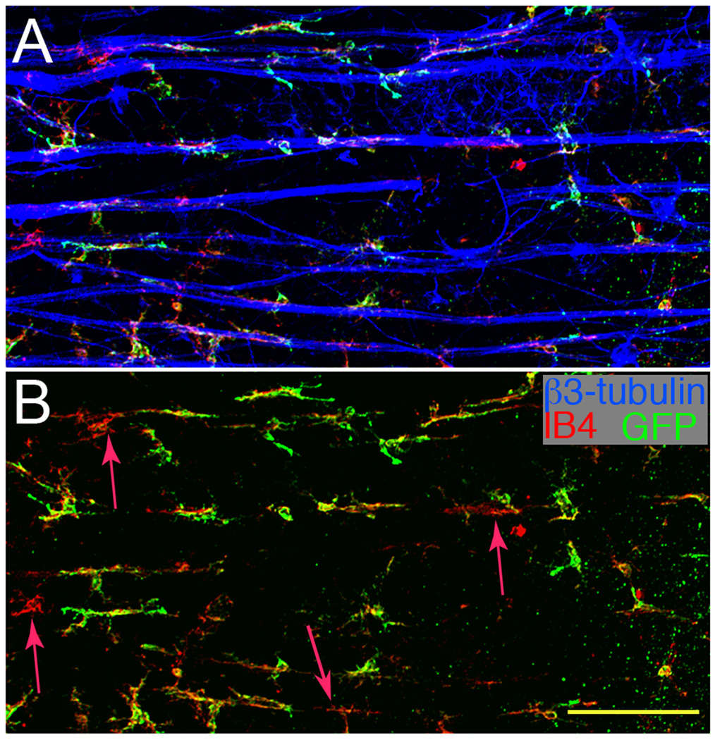
Fig. 6.
The majority of the cells associated with nerve fibers are GFP+CD11b+. (A) A three micron thick optical section of the NFL/RGC from a CD11c-DTR mouse 7 d post-ONC stained for GFP+ cells (green), CD11b+ cells (red), and nerve fibers (blue). (B) Same field as A analyzed for GFP+CD11b+ cells showing very few GFP−CD11b+ cells. Red arrows mark the four cells near nerve fibers that were CD11b+ but GFP−. Scale bar, 100 µm.

